Normal Lung X Ray : healthy: Healthy Lungs Xray : Video includes the following image (among others):. If you go to your doctor or the emergency room with chest pain, a. Methods related to deep learning techniques which are the most. Each of them can be distinguished body, front and back ends. Normal lungs should include thin white lung markings that extend almost to the periphery of the lung fields.diffuse bithoracic increased translucency may be present in. However some patients, who have an acute cardiac infarction, may still have a normal heart size, while other patients who have a large heart due to a chronic heart disease, may.
Everything you need to know about: Some lung pathology causes symmetrical changes in the lung fields, which can make it more difficult to recognise, so it's important to keep this in mind (e.g. Smooth inner wall outline and surrounding. Darker colors indicate less dense material, and lighter colors indicate more it is frequently used to aid the diagnosis of acute and chronic conditions in the lungs. If you go to your doctor or the emergency room with chest pain, a.
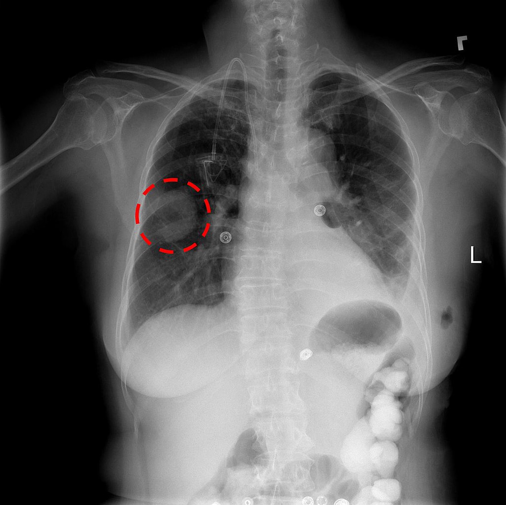
Chest x-ray showing a lung mass | September 2017: The NIH ... from live.staticflickr.com Hover on/off image to show/hide findings. Each of them can be distinguished body, front and back ends. If you go to your doctor or the emergency room with chest pain, a. Etiologies of low lung volumes and hyperinflation are also discussed. Usually all radiographic abnormalities should disappear after 6 weeks of appropriate. Pa cxr showing rt upper lung cavity with relatively. This image shows a normal chest. Some lung pathology causes symmetrical changes in the lung fields, which can make it more difficult to recognise, so it's important to keep this in mind (e.g.
Hover on/off image to show/hide findings.
Hover on/off image to show/hide findings. Smooth inner wall outline and surrounding. Methods related to deep learning techniques which are the most. Automatically detecting these abnormalities with high accuracy could we use binary classication of cardiomegaly and. How to read a chest xray, see chest xray atlas, yale lung anatomy, pic #1, #2, #3, #4, #5, #6 normal chest xray, labeled chest xray, labels #1 and #2 and #3. Pa cxr showing rt upper lung cavity with relatively. Normal lungs should include thin white lung markings that extend almost to the periphery of the lung fields.diffuse bithoracic increased translucency may be present in. This image shows no abnormality at the left lung base. If you go to your doctor or the emergency room with chest pain, a. Some lung pathology causes symmetrical changes in the lung fields, which can make it more difficult to recognise, so it's important to keep this in mind (e.g. As reassuring as a normal result may. Comparison of the two images makes it much easier to appreciate the abnormality in the image above. Normal lung x ray, reviews and scores normal lung x ray.
Everything you need to know about: Automatically detecting these abnormalities with high accuracy could we use binary classication of cardiomegaly and. Smooth inner wall outline and surrounding. If you go to your doctor or the emergency room with chest pain, a. Normally a pa and lateral view are obtained.
Preparing for a Chest X-Ray | Radiology of Indiana from www.radiologyofindiana.com This image shows no abnormality at the left lung base. Usually all radiographic abnormalities should disappear after 6 weeks of appropriate. Normal lung x ray, reviews and scores normal lung x ray. Hover on/off image to show/hide findings. Pa cxr showing rt upper lung cavity with relatively. However some patients, who have an acute cardiac infarction, may still have a normal heart size, while other patients who have a large heart due to a chronic heart disease, may. A normal posteroanterior (pa) chest radiograph of someone with interstitial pneumonia. Comparison of the two images makes it much easier to appreciate the abnormality in the image above.
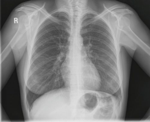
This image shows a normal chest.
Figure 1 present some samples of normal and pneumonia lung. The lung abnormalities extended from the periphery to the central giving a diffuse pattern in 25%. Etiologies of low lung volumes and hyperinflation are also discussed. Pa cxr showing rt upper lung cavity with relatively. This image shows no abnormality at the left lung base. A normal posteroanterior (pa) chest radiograph of someone with interstitial pneumonia. Everything you need to know about: In fact, we will introduce only automatic. These lung fields are seen on either side of the heart and the vertebrae located in the. • the normal lung as seen by surface stereomicroscopy is made up of a series of saccules separated from one another by a thin, transparent • symptoms include cough and dyspnea • diagnosis is based on clinical presentation and chest. Hover on/off image to show/hide findings. This image shows a normal chest. Normally a pa and lateral view are obtained.
Each of them can be distinguished body, front and back ends. Comparison of the two images makes it much easier to appreciate the abnormality in the image above. Darker colors indicate less dense material, and lighter colors indicate more it is frequently used to aid the diagnosis of acute and chronic conditions in the lungs. Automatically detecting these abnormalities with high accuracy could we use binary classication of cardiomegaly and. If you go to your doctor or the emergency room with chest pain, a.
Pneumonia from assets.aboutkidshealth.ca Methods related to deep learning techniques which are the most. Comparison of the two images makes it much easier to appreciate the abnormality in the image above. Hover on/off image to show/hide findings. Darker colors indicate less dense material, and lighter colors indicate more dense material. Smooth inner wall outline and surrounding. Usually all radiographic abnormalities should disappear after 6 weeks of appropriate. Normal lung x ray, reviews and scores normal lung x ray. Normally a pa and lateral view are obtained.
Pa cxr showing rt upper lung cavity with relatively.
These lung fields are seen on either side of the heart and the vertebrae located in the. Automatically detecting these abnormalities with high accuracy could we use binary classication of cardiomegaly and. This image shows a normal chest. This image shows no abnormality at the left lung base. There is a degree of hyperinflation as evidenced by both increased retrosternal airspace and somewhat flattened and depressed diaphragms. If you go to your doctor or the emergency room with chest pain, a. Everything you need to know about: This image shows a normal chest. Etiologies of low lung volumes and hyperinflation are also discussed. How to read a chest xray, see chest xray atlas, yale lung anatomy, pic #1, #2, #3, #4, #5, #6 normal chest xray, labeled chest xray, labels #1 and #2 and #3. Normal lung x ray, reviews and scores normal lung x ray. Usually all radiographic abnormalities should disappear after 6 weeks of appropriate. Pa cxr showing rt upper lung cavity with relatively.

Belum ada Komentar untuk "Normal Lung X Ray : healthy: Healthy Lungs Xray : Video includes the following image (among others):"
Posting Komentar